glaucoma surgery

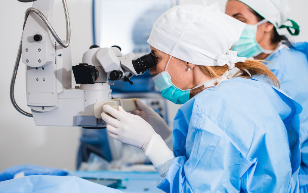
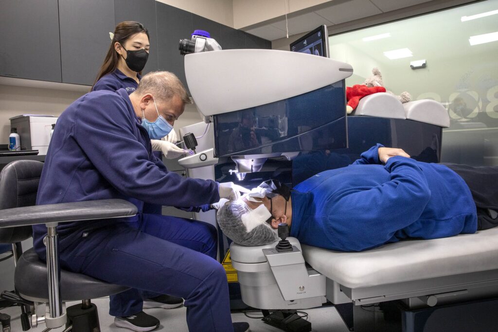
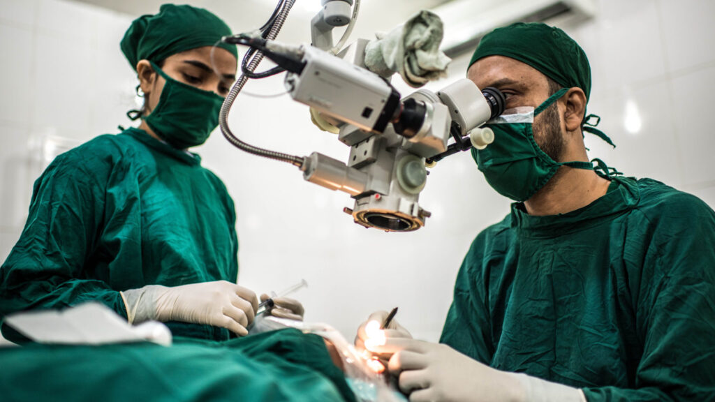
Protecting Your Vision: Glaucoma Surgery in the Context of Retinal Diseases

Glaucoma is a serious eye condition that affects millions of people worldwide. It can lead to vision loss and even blindness if left untreated. In addition to glaucoma, there are several retinal diseases that can also impact your vision. Understanding the connection between these two conditions is crucial in protecting your vision and exploring treatment options such as glaucoma surgery.

Understanding Glaucoma and Retinal Diseases

The eye is a complex organ, comprised of several structures working together to provide visual perception. To better understand glaucoma and its relationship with retinal diseases, it is important to grasp the anatomy of the eye and the functions of different components.

When it comes to managing vision loss caused by glaucoma and retinal diseases, surgery can be a viable treatment option. Glaucoma surgery aims to lower intraocular pressure and preserve vision by improving the drainage of fluid within the eye.

The Anatomy of the Eye

The eye consists of various parts, including the cornea, iris, lens, retina, and optic nerve. The cornea acts as a protective layer at the front of the eye, while the iris controls the amount of light that enters the eye. The lens focuses light onto the retina, which is a thin layer of tissue located at the back of the eye. The retina contains specialized cells called photoreceptors, specifically rods and cones, which convert light into electrical signals that are then sent to the brain via the optic nerve. This intricate process is essential for vision and allows us to perceive the world around us in vibrant detail.

Moreover, the retina is not just a passive receiver of light; it also plays a crucial role in visual processing. The macula, a small but highly sensitive area in the center of the retina, is responsible for detailed central vision and color perception. This region is vital for tasks such as reading, driving, and recognizing faces, highlighting the significance of retinal health in maintaining clear eyesight.

glaucoma surgery

What is Glaucoma?

Glaucoma is a group of eye diseases that damage the optic nerve, often due to increased pressure in the eye. This increased pressure, known as intraocular pressure, can occur when the fluid inside the eye does not drain properly. As a result, the optic nerve becomes damaged, leading to vision loss. Glaucoma is often referred to as the “silent thief of sight” because it can progress slowly and without noticeable symptoms until significant vision loss has occurred. Regular eye exams are crucial for early detection and management of this condition.

Common Retinal Diseases

In addition to glaucoma, there are various retinal diseases that can affect your vision. Some common retinal diseases include age-related macular degeneration (AMD), diabetic retinopathy, and retinal detachment. Age-related macular degeneration is a leading cause of vision loss in older adults, affecting the central vision and making activities like reading and driving challenging. Diabetic retinopathy, on the other hand, is a complication of diabetes that can cause damage to the blood vessels in the retina, leading to vision impairment. Retinal detachment is a serious condition where the retina pulls away from its normal position, requiring prompt medical attention to prevent permanent vision loss. Read more about diabetic retinopathy on https://www.ncbi.nlm.nih.gov/books/NBK560805/

The Connection Between Glaucoma and Retinal Diseases

While glaucoma primarily affects the optic nerve, it can also impact the retina, which is responsible for capturing and processing visual information. The relationship between glaucoma and retinal diseases is complex and multifaceted.

Glaucoma causes damage to the optic nerve, but it can also lead to changes in the retina. As the optic nerve is responsible for transmitting information from the retina to the brain, any damage to the optic nerve can disrupt this flow of information and result in vision impairment.

Furthermore, the impact of glaucoma on the retina can manifest in various ways. In some cases, glaucoma can cause thinning of the retinal nerve fiber layer, which can be detected through specialized imaging techniques like optical coherence tomography (OCT). This thinning of the nerve fiber layer can result in visual field defects, affecting peripheral vision and potentially progressing to central vision loss over time.

Retinal Diseases that can Lead to Glaucoma

On the other hand, certain retinal diseases can increase the risk of developing glaucoma. For instance, conditions like diabetic retinopathy and retinal detachment can lead to changes in eye fluid dynamics, causing an increase in intraocular pressure and subsequently increasing the likelihood of glaucoma development.

Diabetic retinopathy, a common complication of diabetes, can result in the growth of abnormal blood vessels in the retina. These abnormal blood vessels can lead to neovascular glaucoma, a type of secondary glaucoma characterized by the formation of new, fragile blood vessels on the iris that block the normal flow of eye fluid. This blockage can increase intraocular pressure, contributing to optic nerve damage and vision loss if left untreated. To read more about fragile blood vessels click here.

The Importance of Early Detection

Early detection of both glaucoma and retinal diseases is crucial in preventing further damage to your vision. Being aware of the symptoms associated with these conditions and regularly visiting an eye care professional can help catch any issues at an early stage.

Glaucoma is often referred to as the “silent thief of sight” because it can progress without noticeable symptoms until significant vision loss has occurred. On the other hand, retinal diseases like age-related macular degeneration (AMD) can cause a gradual loss of central vision over time. By detecting these conditions early, treatment options can be more effective in preserving your eyesight.

Symptoms to Watch Out For

While glaucoma and retinal diseases can often progress silently, there are some common symptoms to watch out for. These may include blurred vision, difficulty seeing in low light conditions, blind spots in the visual field, and sudden changes in vision. If you experience any of these symptoms, it is important to seek immediate medical attention.

In addition to the symptoms mentioned above, individuals with glaucoma may also experience eye pain, headaches, nausea, and halos around lights. Retinal diseases, on the other hand, can manifest as distortions in straight lines or a decrease in color perception. Recognizing these signs early on can lead to prompt diagnosis and management of these sight-threatening conditions.

Regular Eye Examinations and Their Role

Regular eye examinations play a vital role in the early detection of eye conditions. During these exams, your eye care professional will evaluate the health of your retina and optic nerve, as well as measure your intraocular pressure. These routine screenings can help identify any signs of glaucoma or other retinal diseases before they progress.

Furthermore, eye examinations can also detect systemic conditions such as diabetes and hypertension, which may have ocular manifestations. By monitoring your eye health regularly, you not only safeguard your vision but also contribute to the overall assessment of your well-being.

Glaucoma Surgery as a Treatment Option

Glaucoma is a group of eye conditions that damage the optic nerve, often due to increased pressure in the eye. If left untreated, glaucoma can lead to irreversible vision loss. Surgery becomes necessary when other treatments, such as eye drops or laser therapy, are not effective in controlling the progression of the disease.

glaucoma surgery

Different Types of Glaucoma Surgery

There are various types of glaucoma surgery, each designed to address specific aspects of the condition. Some common procedures include trabeculectomy, in which a small drainage channel is created to reduce intraocular pressure, and tube shunt surgery, which involves implanting a tiny tube to redirect fluid flow. Learn more advanced glaucoma technique on: Advanced Glaucoma Surgery Techniques

In addition to these procedures, newer minimally invasive glaucoma surgeries (MIGS) have been developed in recent years. MIGS techniques use microscopic-sized equipment and tiny incisions to reduce intraocular pressure with fewer complications and faster recovery times compared to traditional surgeries.

The Process of Glaucoma Surgery

Glaucoma surgery typically involves several steps. First, the eye is numbed with local anesthesia. Then, an incision is made to access the drainage structures. The surgeon will then perform the necessary modifications to improve fluid outflow. Finally, the incision is closed, and the eye is allowed to heal.

After surgery, patients may experience some discomfort, blurred vision, or sensitivity to light. It is essential to follow post-operative care instructions provided by the ophthalmologist to ensure proper healing and optimal results.

Risks and Benefits of Glaucoma Surgery

As with any surgical procedure, glaucoma surgery carries certain risks and benefits. While the surgery can effectively lower intraocular pressure and slow down the progression of glaucoma and retinal diseases, there is also the possibility of complications such as infection, bleeding, and inflammation. It is important to discuss the potential risks and benefits with your eye care professional before making a decision.

Ultimately, the goal of glaucoma surgery is to preserve vision and improve the quality of life for individuals affected by this sight-threatening condition. By exploring all treatment options and working closely with your eye care team, you can make informed decisions about the best course of action for managing your glaucoma effectively.

Post-Surgery Care and Management

After undergoing glaucoma surgery, proper post-operative care and management are essential for a successful recovery and long-term maintenance of vision.

Ensuring that you follow your surgeon’s post-operative care instructions diligently can significantly impact the outcome of your surgery. This includes properly administering any prescribed medications, adhering to the recommended schedule for eye drops, and being mindful of any restrictions on activities during the initial healing phase. By taking these measures seriously, you are actively contributing to the success of your recovery process and the preservation of your eyesight.

What to Expect After Glaucoma Surgery

After glaucoma surgery, your eye may be covered with a protective shield or bandage for a short period. You may experience some discomfort, mild pain, or temporary blurred vision. It is important to follow your surgeon’s instructions regarding medication, eye drops, and any necessary restrictions on activities during the healing process.

Additionally, it is common to experience some redness or swelling in the eye following surgery. This is a normal part of the healing process and should subside over time. If you have any concerns about the level of pain or discomfort you are experiencing, do not hesitate to contact your healthcare provider for guidance and reassurance.

Long-Term Management and Follow-Up

Following glaucoma surgery, regular follow-up appointments with your eye care professional are crucial. These visits allow your surgeon to monitor your eye health, assess the success of the surgery, and make any necessary adjustments to your treatment plan. It is important to attend these appointments and communicate any changes or concerns regarding your vision.

Furthermore, maintaining a healthy lifestyle can also contribute to the long-term success of your glaucoma surgery. Factors such as a balanced diet, regular exercise, and avoiding smoking can positively impact your overall eye health and potentially slow down the progression of the disease. By incorporating these habits into your daily routine, you are actively supporting the effectiveness of your treatment and the well-being of your eyes.

Protecting your vision is of utmost importance, especially when dealing with conditions like glaucoma and retinal diseases. By understanding the connection between these conditions and exploring treatment options such as glaucoma surgery, you can take proactive steps towards preserving your vision and maintaining a healthy lifestyle. Remember to prioritize regular eye examinations and seek professional advice for personalized recommendations and guidance.

glaucoma treatment

Retinal Specialist Insights: Advanced Glaucoma Surgery Techniques

Glaucoma is a complex eye condition that affects millions of people worldwide. As a retinal specialist, understanding the intricacies of glaucoma and the latest surgical techniques is crucial in providing the best possible care for patients. In this article, we will explore advanced glaucoma surgery techniques and delve into the role retinal specialists play in the treatment of this sight-threatening disease.

Understanding Glaucoma: A Brief Overview And Treatment

When devising glaucoma treatment plans for glaucoma patients, retinal specialists take a personalized and tailored approach that accounts for the individual characteristics of each case. Factors such as the patient’s age, general health condition, and the progression of the disease are carefully evaluated to determine the most suitable course of action.

The Role of the Retina in Vision

Before delving into the specifics of glaucoma, it is important to understand the role of the retina in vision. The retina is a complex and vital component of the eye, serving as a thin layer of tissue located at the back of the eye. It contains millions of specialized cells called photoreceptors that convert light into electrical signals, which are then transmitted to the brain through the optic nerve.

These photoreceptors, known as rods and cones, play a crucial role in our ability to see and interpret the world around us. Rods are responsible for vision in low light conditions and help us see shapes and movement, while cones are essential for color vision and detailed central vision. The intricate workings of the retina allow us to perceive the rich tapestry of colors, shapes, and textures that make up our visual experience.

Without a healthy retina, our vision is compromised, and this is where glaucoma poses a significant threat. Glaucoma is a group of eye conditions that damage the optic nerve, often as a result of increased pressure within the eye. If left untreated, glaucoma can lead to permanent vision loss.

What is Glaucoma and How Does it Affect Vision?

Glaucoma is often referred to as the “silent thief of sight” because it typically progresses slowly and without obvious symptoms in its early stages. As the disease progresses, however, individuals may experience peripheral vision loss, tunnel vision, or even complete vision loss in severe cases.

The exact causes of glaucoma are still being studied, but high intraocular pressure (IOP) is a significant risk factor. Normally, fluid called aqueous humor circulates within the front portion of the eye. In individuals with glaucoma, this fluid does not drain properly, leading to increased pressure within the eye.

Early detection and treatment are crucial in managing glaucoma and preserving vision. Regular eye exams, including measurement of intraocular pressure and evaluation of the optic nerve, are essential in detecting glaucoma in its early stages. Treatment options may include eye drops, oral medications, laser therapy, or surgery, depending on the type and severity of the condition.

glaucoma treatment

The Evolution of Glaucoma Surgery Techniques

Traditional Glaucoma Surgery Techniques

In the past, traditional glaucoma surgery techniques aimed to lower intraocular pressure (IOP) through surgical procedures like trabeculectomy or implanting drainage devices. These procedures were effective but often came with risks and complications.

Trabeculectomy involves creating a new drainage channel for aqueous humor to lower IOP. While effective, it can lead to complications such as infection or scarring. Similarly, implanting drainage devices can be successful, but the risk of infection and the need for periodic adjustments pose challenges. Read more about drainage channel at https://www2.tulane.edu/~sanelson/eens1110/streams.htm

The Shift Towards Advanced Techniques

Advancements in technology and surgical techniques have led to the development of more advanced glaucoma surgery options. These advanced techniques aim to provide better outcomes with fewer complications for patients.

A notable example of advanced glaucoma surgery is Laser Trabeculoplasty. This procedure uses a laser to target the drainage angle of the eye, enhancing fluid outflow and reducing IOP. Laser Trabeculoplasty is a minimally invasive option that can be performed in an outpatient setting, offering advantages such as shorter recovery times and reduced risk of complications.

Another advanced technique gaining popularity is the use of Ex-PRESS mini shunt. This device is a small stainless steel tube that is inserted into the eye to create a controlled drainage pathway. The Ex-PRESS mini shunt has shown promising results in reducing IOP and minimizing the need for postoperative interventions.

Minimally Invasive Glaucoma Surgery (MIGS)

Another category of advanced glaucoma surgery techniques is Minimally Invasive Glaucoma Surgery (MIGS). MIGS procedures involve the use of small incisions to implant microstents or other devices that improve aqueous outflow and reduce IOP.

These MIGS procedures offer several benefits, including decreased surgical trauma, reduced dependency on glaucoma medications, and faster recovery times. While not suitable for all glaucoma cases, MIGS has become a valuable addition to the glaucoma surgeon’s armamentarium.

One of the innovative MIGS procedures is the iStent. This tiny titanium device is implanted into the eye’s drainage system to improve the outflow of aqueous humor. The iStent has shown promising results in reducing IOP and decreasing the need for glaucoma medications.

Trabeculectomy and Tube Shunt Surgery

Despite the advancements in advanced glaucoma surgery techniques, traditional procedures like trabeculectomy and tube shunt surgery still play a significant role in the management of more severe or complex cases of glaucoma.

Trabeculectomy involves creating a small flap in the sclera (the white part of the eye) to create a new drainage channel. Tube shunt surgery, on the other hand, involves implanting a small tube in the eye to facilitate the drainage of aqueous humor.

While these procedures may carry a slightly higher risk of complications compared to some advanced techniques, they remain effective options for certain patients, particularly those with advanced glaucoma or previous surgical interventions. To read more about surgical interventions click here.

It is worth noting that ongoing research and technological advancements continue to shape the field of glaucoma surgery. Surgeons are constantly exploring new techniques and devices to improve patient outcomes and minimize the burden of glaucoma on individuals’ lives.

The Role of Retinal Specialists in Glaucoma Treatment

The Expertise of Retinal Specialists

Retinal specialists are highly trained ophthalmologists who have pursued additional education and experience in diagnosing and treating diseases that affect the delicate structures of the retina and vitreous. While glaucoma primarily impacts the optic nerve, it can also lead to detrimental effects on the retinal tissue. This is where the specialized skills of retinal specialists come into play, as they are equipped to provide comprehensive care for patients with glaucoma.

With a profound understanding of the intricate anatomy and function of the retina, retinal specialists are uniquely positioned to evaluate the implications of glaucoma on this vital part of the eye. Their expertise allows for a thorough assessment of how glaucoma is affecting the retina, guiding them in making well-informed decisions regarding the most effective treatment strategies. By considering both the health of the optic nerve and the overall function of the retina, retinal specialists ensure a holistic approach to managing glaucoma.

The Retinal Specialist’s Approach to Glaucoma

Moreover, retinal specialists go beyond simply addressing intraocular pressure (IOP) levels in glaucoma management. They also focus on monitoring and addressing any potential complications that may arise from the treatment itself. Through regular examinations and specialized diagnostic tests, retinal specialists track the progress of the disease, ensuring that any changes in the retinal tissue are promptly identified and managed with precision and expertise.

Advanced Glaucoma Surgery Techniques

Laser Trabeculoplasty

Laser Trabeculoplasty is a non-invasive procedure that can be done in an outpatient setting. During the procedure, a laser is used to apply focused energy to the drainage angle of the eye, increasing drainage and reducing intraocular pressure (IOP).

This technique offers several advantages, including minimal discomfort, quick recovery, and the potential to decrease reliance on glaucoma medications. By targeting the drainage angle, Laser Trabeculoplasty helps to improve the flow of aqueous humor, the fluid within the eye, and thus manage glaucoma. While the effects of Laser Trabeculoplasty may not be permanent, it can help delay the need for more invasive surgical interventions.

Imagine a patient, Mr. Johnson, who has been struggling with glaucoma for years. His daily routine involves taking multiple eye drops to manage his condition. Laser Trabeculoplasty offers him a glimmer of hope, as it can potentially reduce his reliance on medication and improve his quality of life. With minimal discomfort during the procedure and a quick recovery period, Mr. Johnson can look forward to a brighter future with better control over his glaucoma.

Minimally Invasive Glaucoma Surgery (MIGS)

Minimally Invasive Glaucoma Surgery (MIGS) encompasses a range of procedures, each with its unique approach. These techniques typically involve creating small incisions or implanting microstents to enhance aqueous outflow and lower IOP.

One such MIGS procedure is the insertion of a tiny stent into Schlemm’s canal, a critical channel involved in aqueous humor drainage. This approach provides a safe and effective means of reducing IOP, often with a minimal impact on the patient’s daily routines. By improving the outflow of aqueous humor, MIGS procedures help to alleviate the pressure within the eye and manage glaucoma.

Let’s consider the case of Mrs. Anderson, a busy professional who is constantly on the go. She has been struggling with glaucoma and finds it challenging to adhere to her medication regimen. MIGS offers her a promising solution, as it can provide a long-term reduction in IOP with minimal disruption to her daily activities. With the insertion of a microstent into Schlemm’s canal, Mrs. Anderson can enjoy improved eye health and a better quality of life.

Trabeculectomy and Tube Shunt Surgery

Trabeculectomy and tube shunt surgery are considered more invasive approaches to glaucoma treatment. These procedures involve creating new drainage channels or implanting tubes to facilitate the outflow of aqueous humor.

While the recovery from these surgeries may be longer, they can be highly effective in managing glaucoma, particularly in cases where other surgical options may not yield sufficient results. Trabeculectomy involves creating a new drainage channel, bypassing the natural drainage system, to reduce IOP. Tube shunt surgery, on the other hand, involves implanting a small tube to redirect the flow of aqueous humor and lower IOP.

Consider the case of Mr. Thompson, who has been battling severe glaucoma for years. Despite trying various treatments, his IOP remains dangerously high, putting his vision at risk. Trabeculectomy or tube shunt surgery may be his best options to regain control over his glaucoma. While the recovery period may be longer, the potential benefits of these procedures, such as a significant reduction in IOP, make them worth considering for patients like Mr. Thompson.

glaucoma treatment

The Benefits and Risks of Advanced Glaucoma Surgery Techniques

Evaluating the Benefits

Advanced glaucoma surgery techniques bring numerous benefits to patients and healthcare providers alike. These techniques often result in lower IOP, thus reducing the risk of further damage to the optic nerve and retinal tissue.

For patients, the advantages include improved vision preservation, reduced reliance on glaucoma medications, and an enhanced quality of life. Additionally, the less invasive nature of advanced techniques leads to shorter recovery times, fewer complications, and an increased likelihood of maintaining overall ocular health.

Understanding the Potential Risks

While advanced glaucoma surgery techniques have significantly improved patient outcomes, it is important to acknowledge that all surgical procedures carry some level of risk. Complications can range from infection and bleeding to vision disturbances and scarring.

However, the benefits of these surgeries often outweigh the risks, particularly in cases where glaucoma progression poses a significant threat to a patient’s vision. Surgeons carefully assess each patient’s unique circumstances and discuss the potential risks and benefits before proceeding with any surgery.

Conclusion

As a retinal specialist, being knowledgeable about advanced glaucoma surgery techniques is essential in providing the best possible care for patients. Glaucoma, a condition that can steal one’s vision if left untreated, requires a multidisciplinary approach that combines the expertise of various eye care specialists.

The evolution of glaucoma surgery techniques has brought about significant advancements, with laser trabeculoplasty and minimally invasive glaucoma surgery (MIGS) offering safer and more effective options for patients. Traditional procedures like trabeculectomy and tube shunt surgery continue to play a crucial role in managing complex cases.

Working alongside ophthalmologists, retinal specialists ensure that glaucoma treatment incorporates a comprehensive understanding of both the optic nerve and the retinal tissue. By considering the benefits and risks of advanced glaucoma surgery techniques, retinal specialists can provide patient-centered care that maximizes vision preservation and enhances the quality of life for individuals living with glaucoma.

Read more about glaucoma surgery on: Glaucoma Surgery in the Context of Retinal Diseases

Getting LASIK Surgery

What Should be the Motivating Factors to Getting LASIK Surgery?

The development of LASIK laser eye treatment in the 1990s has improved the quality of life for millions of people throughout the world. One of the most sought-after corrective laser procedures due to its short procedure time and rapid visual recovery. 

It’s natural for people to ask if LASIK eye surgery is a good option for them, given the procedure’s widespread publicity and high success rate. In this article, we’ll take a deeper look at eye surgery with LASIK to see if it’s the perfect option for you, how it operates, and how it can enhance your life through improved vision.

What is Eye Surgery LASIK?

Eye Surgery LASIK

Laser-Assisted in-situ keratomileusis is more commonly known as LASIK. This surgical treatment reshapes the cornea to improve eyesight and eliminate the need for corrective lenses. It is effective, precise, and carries a negligible risk of problems that are easily remedied and rarely result in permanent vision loss.

Multiple types of refractive defects are amenable to LASIK eye surgery. For instance;

  • Up to +4.00 D of hyperopia (farsightedness)
  • Up to -12.00 D Myopia (nearsightedness)
  • And a 5.00 D of astigmatism with them.

Millions of individuals around the world who have trouble seeing can now look forwards to a brighter future because of this. If you’re considering surgery with LASIK, your doctor will assess if you’re a good candidate during your first consultation. 

Fortunately, if you decide eye surgery using LASIK isn’t for you, there are many more laser eye treatment options available thanks to recent advancements in the field. learn more about laser eye surgery cost by clicking here.

When LASIK is Performed, How Does it Work?

LASIK is Performed

Your LASIK eye doctor will use a Femtosecond Laser to cut a tiny flap in the cornea. In order to correct the specified visual issue, the freshly generated flap is lifted, and a second laser is utilised to remodel the corneal tissue lying beneath. Next, the corneal flap is repositioned.

The ultimate effect is a cornea that better focuses light on the retina, resulting in sharper vision. Even though most patients have a sudden and significant improvement in their eyesight, it continues to enhance over the next few weeks.

Is LASIK a Good Idea for You?

Here are some pointers to consider as you weigh the benefits and drawbacks of LASIK eye surgery in light of our newfound knowledge.

LASIK eye surgery is something to think about if you:

Need to Achieve Complete Visual Independence in One Day

Fast recuperation time is a major selling point for LASIK eye surgery. Although there are a number of approaches to addressing vision problems, very few of them offer quick fixes that last. Some people are interested in the possibility of having their vision improved but are not yet prepared to undergo the laser eye treatment. 

However, most eye doctors will be ready to schedule your LASIK surgery right away. In addition, you might be able to ditch your corrective lenses altogether after surgery.

Have negative experiences with contacts or glasses

You should really consider eye surgery with LASIK if you have a strong dislike for either your glasses or your contacts. Glasses and contacts are something that some individuals may love using, while many others learn to despise for various reasons. 

Glasses can pinch, shatter easily, and get in the way when you’re trying to be active or do particular things, which are all reasons why many people choose not to wear them. However, contact lenses can have negative side effects, including dry eyes and other irritations. 

In addition, they all have the potential to induce nausea, vomiting, or dizziness. But with LASIK eye surgery, most people never need to wear corrective lenses again.

Health Related Problems

Similarly, if you’re worried about your health, LASIK eye surgery could be a good investment. For those who suffer from vertigo, migraines, and other conditions that might be exacerbated by wearing glasses, this is a real concern. 

Furthermore, wearing contacts can be very unpleasant if you have allergies or any other eye-related concerns. This is due to the fact that exposure to them might result in symptoms such as dryness, redness, irritation, itching, ocular infections, and oxygen deprivation. Putting contacts in eyes that are already having problems can exacerbate the condition.

Booster of Confidence

Booster of Confidence

Some people feel more confident when they wear glasses, but for others it’s the opposite. If you fall into the second category, LASIK eye surgery is a rapid and effective solution. Instead of being shackled by glasses that make you feel less than your best, LASIK surgery could be your ticket to freedom and a positive self-image.

Athletics

Some sports and other physical pursuits are inaccessible to people who must wear glasses. This is due to the fact that glasses are prone to breaking in the midst of any sort of vigourous exercise. Payment for specialist (and typically quite costly) athletic eyewear is one solution. 

Instead, you can try hiding them inside a (often very unattractive) protective cover, which won’t necessarily keep your glasses from slipping off or breaking even if you’re careful. But if you have eye surgery with LASIK, you won’t have to worry about breaking your glasses or hurting your eyes while playing sports because the surgical procedure eliminates the need for corrective lenses.

Work Responsibility

If you work in an environment where glasses or contacts aren’t allowed, that’s another plus for surgery with LASIK. Many law enforcement-related institutions and programmes exclude people with visual impairments, including police forces, fire departments, and the armed services. In order to avoid missing out on your ideal career because of vision problems, LASIK eye surgery is a fast and simple option to consider.

Constant Movement

Finally, your glasses or contacts might be starting to bother you if you’re a frequent flyer. The more you travel, the more likely it is that you may lose, misplace, or damage your glasses or contacts. There are a lot of places in an airport or railway station, hotel room, or restroom where you could accidentally set down your glasses case or remove your contacts and lose them forever. If you spend the money on eye surgery involving LASIK, you won’t have to worry about your eyesight again.

Contact Personal Eyes Today to Schedule Your FREE LASIK Consultation

It’s safe to say that you’re not alone if LASIK surgery is something you’re considering. As many as a million people have already had this laser eye treatment done, and another million are just waiting for the proper time to schedule it. 

LASIK surgery can be the ticket to visual independence for anyone with blurred vision who values mobility, athleticism, health, tired of glasses or wear contact lenses, or the pursuit of a new job but is limited by their eyesight. However, because each person’s situation and outcomes are unique, you should carefully weigh the benefits and drawbacks of the procedure before deciding to go through with it.

Personal Eyes’ mission is to provide information to everyone interested in learning how to better their eyesight. And if you are seriously considering laser vision correction, you need first ensure that you are a suitable candidate.

If you are having any question or inquiry about how to have a correct vision. Laser eye surgery procedure, Corneal tissue problems, Lasik surgery, laser beam in healing process, excimer laser usage, refractive eye surgery, laser treatment, laser suite, contact sports, the surgical procedure for refractive surgery or Lasik laser eye surgery – you should get in touch with us via our Personal Eye website to book your free assessment today.

sydney eye clinic

Common Questions About Diabetes and Eye Surgery: Sydney Eye Clinic

What are the risks of having cataract surgery if I have diabetes?

In most cases, patients with diabetes can safely undergo any kind of eye surgery. However, your overall health and absence of any other medical concerns will determine whether or not you are a candidate for eye surgery.

In rare situations, your overall health or other medical concerns may need you to postpone eye surgery. To find out if you are a candidate for eye surgery and when it would be best for you to have the procedure, consult with an ophthalmologist.

Prior to any major surgical procedure, you will likely have an appointment with sydney eye clinic or ophthalmologist to make sure your diabetes is not affecting your eyesight. 

First, you’ll want to get your eyes checked by an optometrist for a quick check-up, and then you may visit an ophthalmologist for a more thorough examination. Additional care will be discussed and planned for in the weeks leading up to your scheduled eye surgery.

All of the medical professionals working on you, from the ophthalmologist to the anaesthesiologist to the surgical nurses and the clinical team, are well-versed in caring for patients with diabetes and are committed to ensuring that you have the best possible surgical outcome. Care of this calibre is provided before, during, and after operation.

We have taken due diligence to publish articles such as this to help you live a healthy life. Our Personal Eye doctors are professionals who create medium for eye health through laser vision correction, various laser treatment and surgeries in New South Wales and the rest of Australia. 

Eye patients have been choosing eye drops, intravitreal injections, and surgeries such as pterygium surgery, LASIK surgery and other latest technology for their eye conditions. we hope that you will find this post about home techniques to manage itchy eyes, which was written by our refractive surgeons, resourceful.

Related: Considering Eye Surgery? LASIK May Be the Right Option for You

Does my HbA1c or blood sugar need to be under control before surgery?

Blood sugar level (BSL) levels tend to differ from patient to patient since diabetes is a complicated condition. Because of this, you should learn the proper methods for managing your diabetes from both your primary care physician and endocrinologist; there is no “one size fits all” solution.

If you want to keep your diabetes under control as much as possible, you may need to keep your average BSL or HbA1c level within a certain range. When you’re inside this range, you’re at peak health and safety for any medical or surgical operation, including eye surgery.

There is a low chance of problems after standard eye surgeries. To further improve your chances of a successful surgical outcome, however, you should ensure that your blood sugar and/or HbA1c levels are within the ‘safe’ range before the procedure.

Your ophthalmologist will coordinate with your primary care physician and endocrinologist to get you ready for surgery.

When I have my eyes fixed, do I have to fast?

Before undergoing eye surgery, patients are often told to abstain from all food and liquids. This is because some patients have a temporary loss of reflexes after receiving sedatives from their surgeon prior to an eye operation.

There is always the risk of choking or vomiting if there is food or liquid in your stomach. If this occurs, the food particles may go to the lungs, where they may obstruct or even damage the airways.

Talk to your eye surgeon ahead of time about your requirement for regular meals and fluids if you have diabetes. For this reason, diabetic patients are generally put at the front of the line for scheduled eye surgeries.

Should I stop taking my medicine before having surgery on my eyes?

Your ophthalmologist has to know about any and all medications you are taking, not only those for diabetes. It all depends on the sort of operation you’re having, but you may be asked to cease taking your medicine. If you’re having eye surgery, for example, you could be told to cease taking your blood thinners.

This is something that you and your treating doctors will need to address. You may feel confident that you will get detailed, written instructions well in advance of your operation if you need to make any adjustments to your regular prescriptions.

In order to have the most successful outcome from my upcoming eye surgery, what should I do in advance?

Before undergoing eye surgery, it’s advisable to follow the pre-op BSL optimisation recommendations of both your ophthalmologist and primary care physician/endocrinologist. The better your chances of having a successful operation and recovery, the longer your BSL stays within the recommended range for you (as determined with your doctor).

What can I expect if I have diabetes and need surgery?

After the procedure, you should be able to see beautifully. It’s possible that diabetic patients‘ “follow-up pathway” is modified slightly.

Your ophthalmologist may recommend more regular check- ups after surgery to check for any complications caused by your diabetes and its possible impact on your eyesight. You may end up needing to see an eye doctor more often than you anticipated, but this is temporary and will ultimately be to your benefit.

During my recuperation, what should I anticipate?

Recuperation following eye surgery often isn’t affected by diabetes. However, it’s possible that you’ll go through the typical recuperation hiccups. After having cataract surgery, for instance, many people report feeling like there’s something gritty in their eye.

Eyelid cleanliness, as instructed by your optometrist and/or ophthalmologist, and the postoperative eye drops supplied to you should help with this.

It’s vital that you report any unusual symptoms to your eye care team so they can be looked at. During your follow-up appointments after eye surgery, every member of your eye care team will assess your progress and provide recommendations for how to speed up your recovery.

Be sure to take it easy for at least a week after surgery. This includes putting aside time for things like golf, gardening, and swimming. Sunglasses are essential for protecting your retina from harmful UV rays throughout the healing process and should be worn at all times outside.

Your current glasses will work fine for reading and watching television. The likelihood of a prescription changes a month following surgery is high.

Maintain your usual routine of testing your blood sugar levels (BSL) and taking your diabetic medicines as directed by your primary care physician or endocrinologist.

Lastly

If you are having any question or inquiry about how to have a correct vision, cataract surgery, Laser eye surgery, laser vision correction, diabetic retinopathy, minimally invasive glaucoma surgery, normally clear lens, vision loss, retinal conditions, short sightedness, – you should get in touch with us via our Personal Eye website to book your free assessment today.

Consider the factors outlined in this blog post, weigh the pros and cons, and most importantly, consult with a Sydney cataract surgeon at Personal Eyes before making the final decision.

Multiple Sclerosis

Multiple Sclerosis: A Revival of Hope

Imagine watching a woman with multiple sclerosis of many years duration (who had previously needed a “walker” to help her get around) walking unaided several times around a room at good speed and with no balance problems. Imagine listening to her say she’s sleeping better, her energy is much improved, and that she’s able to think more clearly. She attributes her dramatic improvement to the natural amino-acid derivative she’s been using for the previous two to three weeks. Imagine hearing another woman, much more seriously afflicted, report that she’s able to feed herself again, and that her friends and relatives had all noticed her speech is easier to understand. Both of these improvements occurred within a month of starting the “new” natural amino-acid derivative.

Your editor has seen and listened to both these women in just the last month. One of your editor’s colleagues at Tahoma Clinic, Dr. George Gillson, M.D., Ph.D., reports that at first checkback (approximately six weeks for treatment) for nineteen individuals with multiple sclerosis, eleven noted dramatic improvement, three reported one or more significant improvements in symptoms (including reduced numbness, better motor control, improved speech, much better sleeping, and more energy) one had no change, and four had no change associated with poor absorption of the natural amino-acid derivative, poor patch adhesion, or an interfering drug.

The nurse responsible for the revival of the use of the natural amino-acid derivative (a now mostly symptom-free “MS” sufferer herself) has collected verbal reports from over 200 individuals diagnosed with “MS” who’ve used the natural amino-acid derivative: 72% report at least one significant improvement in symptoms, and some many more.

The Natural Amino Acid Derivative: 

The natural amino acid-derivative is histamine, a very small and simple molecule made by every human (and animal) body from the naturally occurring (and conditionally essential) amino-acid histidine. Yes, that’s the same histamine that most of us are told is the “bad guy” of the allergy world, against which we’re all urged to swallow the latest patent (and prescription-only, until the patent expires) “antihistamine” medication. Apparently, individuals with “MS” either don’t make enough histamine in their own bodies, or just need more. Perhaps both. No one knows for certain.

Isn’t it premature to be writing about a symptom improvement in MS based on verbal reports of only 200+ individuals, and only 19 reporting back so far to Tahoma Clinic? Results achieved with… of all things… histamine? Isn’t is all too new…and perhaps too wacky…to get our hopes up? Please refer back to title of this report (A Revival of Hope), and then let’s travel back in time to St. Joseph’s Hospital in Tacoma, Washington. The year is 1950; the reporter is Miriam Zeller Gross, who published the article from which the following paragraphs are excerpted from McCall’s Magazine for December of that year.

“Take Mrs. Alice Meinert. This young mother was stricken shortly after New Year’s Day in 1947. By May, she could not get out of bed. For a year she grew steadily worse. Her father heard about the Clinic in Tacoma [not Tahoma Clinic, which was founded in 1973.-ed.]. He urged that his daughter be sent there. But the attending physician pooh-poohed the idea, and acting on his advice, Mrs. Meinert’s husband refused.

“The father took legal action and gained custody of his daughter- a step accomplished through the farseeing wisdom of Judge Chester A. Batchelor of the King County Court, Seattle.

“Four days after she reached the Tacoma clinic, Mrs. Meinert took her first steps in more than a year. One week later she walked from the house to the street and got into an automobile unaided…..her condition has improved steadily. She does her own housework, looks after her child, and appears in every way to be a well, happy woman.”

See also: You’re just 24 hours away from discovering-and reducing-your breast cancer risk

Multiple Sclerosis

Dr. Hinton Jonez: 

In 1946, Hinton Jonez, M.D., a Tacoma general practitioner, was invited by the Sisters of St. Joseph’s Hospital to open an MS clinic in a hospital wing. The Sisters had observed improvements in several individuals with MS hospitalized at St. Joseph’s under Dr. Jonez care. They had observed that the mainstay of Dr. Jonez’ treatment was injectable histamine, and knew that injectable histamine could cause adverse effects, including severe headaches or stomach aches with considerable cramping if injected at the wrong dose or speed of administration. There were even reports of deaths from too much histamine injected too rapidly. But Dr. Jonez’ patients had had no such adverse effects, and all had improved, so the Sisters were happy when Dr. Jonez volunteered to open a clinic at St. Joseph’s dedicated to the treatment of MS.

Dr. Jonez had learned of injectable histamine treatment for MS at a meeting of the American College of Allergy (now called the American College of Allergy and Immunology) from the then-well-known Bayard T. Horton, M.D., of the Mayo Clinic (Rochester, Minnesota). According to Dr. Jonez, when discussing allergy and allergy treatment over dinner, Dr. Horton had told him and a group of physicians: “Take multiple sclerosis. There is good reason to believe it is an allergic condition. According to Dr. Jonez, Dr. Horton had explained that histamine gives new life to MS victims much as fresh fighting troops revive an exhausted army. “It’s too early to say much,” Dr. Horton told Dr. Jonez, “but we believe we are on the right track.”

Histamine For Allergies: 

Some fifty or more years after Dr. Horton’s time, we’ve all been thoroughly convinced by the patent medicine companies propaganda (“advertising”) that patented and formerly patented “antihistamines” are the best way to combat allergy. Such was not the case in the 1940′s. Dr. Jonez explains:

“Let me review some 1946 medical history…the antihistamine drugs were big news that year…pharmaceutical houses worked night and day to rush the latest and most potent antihistaminic drug to doctors and druggists….while most doctors dosed their patients with antihistamine s, Dr. Horton did the exact opposite. He administered histamine. And he was getting results. Both allergic conditions and an impressive array of illnesses were yielding to Dr. Horton’s histamine treatment.

“Mysterious, intolerable head-aches disappeared. So did the symptoms of Meniere’s disease, characterized by progressive deafness, previously relieved by highly delicate surgery. A host of bizarre eye and ear conditions heretofore thought incurable had also responded to histamine.

“Horton’s method was in a sense fighting fire with fire, and based on the same line of reasoning as giving cowpox vaccinations to fight smallpox….Instead of suppressing the action of histamine by antihistaminics, he used histamine against histamine.”

If these successes were achieved by a respected staff member of the Mayo Clinic in the 1940s, why isn’t histamine commonly in use today against allergic diseases? The answers lie in the nature of histamine itself, and in the nature of American medicine. Histamine is a very “unstable” molecule; it “breaks down” very rapidly. When given orally, it can cause considerable stomach upset and cramping; when given too rapidly or in too great a quantity by injection it can (as noted above) give very unpleasant effects. A few people had actually died after being injected with too much histamine too fast. But administering it properly, Dr. Horton reported that he had given thousands of injections without a single ill effect. So why didn’t physicians learn and apply Dr. Horton’s methods as Dr. Jonez did?

Histamine’s biggest “handicap” is (and was) that as a molecule produced in human bodies it isn’t patentable. Patent medicine (sometimes called “pharmaceutical”) companies would not work “night and day” to rush histamine…or news of its latest uses…to doctors and druggists. And in the 1940s, as now, the vast majority of physicians got most of their “new treatment” information from patent medicine companies.

Dr. Jonez First “Case”: 

Mrs. Johnston had suffered from MS for five years. She was bedridden, unable to move her legs. She was going blind, and had difficulty swallowing.

Dr. Jonez describes her response to histamine treatment:

“[Histamine] was given slowly, carefully. All the elaborate precautions Horton outlined were observed. He had said that histamine had an unwarranted bad reputation because doctors…gave [it] too rapidly, or used contaminated equipment. They failed to realize that the fault lay in their own ineptness….

“A rosy glow spread over Mrs. Johnston’s face, then down the arms. “I feel better already,” she said…As the days went by, there was no doubt she was getting better….she could swallow with ease for the first time in months. And to the amazement of her eye specialist, her vision was back to normal….Less than six months after her first dose of histamine, she was walking. Sensation had fully returned to the legs that had appeared hopelessly paralyzed. It began the evening her husband telephoned in great excitement; “She can wiggle her toes!…The progress was steady. Soon she gave away the wheelchair.”

Dr. Jonez goes on to explain that the natural course of MS can include unexplained “spontaneous” remissions, sometimes of long duration. As this was his first case, he couldn’t be certain that the histamine injections had caused Mrs. Johnston’s improvement. How-ever, five years later, after administering some 150,000 doses and observing the results, he wrote: “…histamine is the medication of first choice in multiple sclerosis.”

Dr. Jonez’ Clinic at St. Joseph’s: 

After obtaining space at St. Joseph’s and with the help of the sisters, Dr. Jonez added several features to the basic histamine treatment. As Dr. Horton had told Dr. Jonez that MS was caused by allergy, and since Dr. Jonez’ use of Dr. Horton’s histamine treatment for MS had been successful in many cases, it’s understandable that Dr. Jonez wrote (in a “professional report”): “At our clinic, complete allergy management is the basis of therapy. On all of our patients, allergy histories are taken and scratch tests [the best available at the time – ed.] are made for sensitivities to foods, epidermals, molds, fungi, pollens, and miscellaneous allergens.” On the basis of these test, diets and allergy desensitization programs were individualized for each MS patient. Dr. Jonez emphasized the importance of allergy control as well as histamine treatment: “Almost without exception, our chronic, progressive [MS patient] suffered from food allergies.” He recounts the case of a patient: “who was out of his wheelchair three times and back again because he thought a small order of salmon and spinach wouldn’t make any difference.”

Physical therapy was another important part of Dr. Jonez’ program. The Sisters of St. Joseph’s helped him to make sure physical therapy was done adequately and appropriately for each patient. For patients whose muscles were twisted and contorted with MS spasm, Dr. Jonez prescribed injections of a powerful muscle relaxant to aid in muscle manipulation.

After five years, Dr. Jonez’ multiple sclerosis program was so successful that the Sisters decided to erect a new clinic building to house what would be named St. Joseph Hospital Clinic for Demyelinating Diseases. The official “groundbreaking” occurred on December 8,1951, with opening scheduled for August 15, 1952. Unfortunately, Dr. Hinton Jonez died, the Sisters could not find even one physician on St. Joseph’s staff willing to continue his program, and Dr. Jonez’ clinic and program at St. Joseph’s in Tacoma came to an end.

Other Natural Treatments for MS: 

At the same time Dr. Jonez was working at St. Joseph’s, another pioneer in effective natural MS treatment, Dr. Roy Swank, was developing his MS diet while on the faculty of the University of Oregon Health Sciences Center. Dr. Swank’s diet is high in “unsaturated fatty acids” which have been found to aid MS when supplemented alone. Others (including Dr. Jonez) were exploring the use of injectable Vitamin B12, as well as injectable adenosine monophosphate (AMP), a natural substance made within every cell of our bodies. During the intervening years, your editor (as well as others) have found that a large proportion of individuals with MS have significant impairments of digestion and assimilation, and that a unique herbal combination can have a significant effect in MS treatment. Your editor and other Tahoma Clinic physicians have observed DHEA to be a small help for some individuals with MS. All of these valuable natural treatments and aspects of MS will follow the description of the “improved histamine” Procarin, the invaluable contribution of Elaine DeLack, R.N.

Elaine DeLack, MS, And Procari: 

Elaine developed her first symptoms of MS in 1985 while living in Montana. While pregnant with her son, she developed difficulty moving her left leg. After delivery, she had variable difficulty moving her left arm and hand. In 1987, a MRI (magnetic resonance image) showed what appeared to be MS lesions in her central nervous system; a second MRI showed more lesions, and the “official diagnosis” of MS was made in 1988.

She continued to worsen until “making dinner was a chore.” Finally, she had her self-described “wake-up call”; A fall while carrying her son, who required stitches for his cuts. She knew she needed more help. She received a telephone call from a caring woman who advised her to call Raymond Bjork, M.D., a Montana physician, who advised her to try injections of Vitamin B12 and adenosine monophosphate (AMP). She reports “it really helped,” and that she could put herself into remission with these natural injections.

In 1993, she finished work for her R.N., which she had started in Montana before taking “time out” to care for her children. While working at her first job at a nursing home, she tried to convince the attending physicians to use injections of Vitamin B12 and AMP for MS patients. Only one physician would listen; he had the injections given to one of the nursing home residents suffering from MS, who strengthened and went home. Despite this none of the other physicians would consent to try the injections for their MS patients, telling Elaine “there isn’t enough research.”

So in 1994, she enrolled in a research-methods course at the Bothell campus of the University of Washington, determined to find and develop research on injectable Vitamin B12 and AMP. She found research showing that histamine stimulated the production of the “intrinsic factor” by the stomach. She knew that Vitamin B12 cannot be absorbed without “intrinsic factor,” and she recalled that Vitamin B12 had not worked for her when she swallowed it, but had been very helpful for her when she injected it. She felt she had found key information. After that, it seemed that in her research “everything led to histamine.”

Elaine started giving herself histamine by injection and with transdermal patches, but found the effects to transient. Further research led her to other natural substances, which would slow the body’s breakdown of histamine. She found a combination which helped her eliminate all her own MS symptoms. After first working with Judy Richardson, R.Ph., who helped develop the delivery system, she located George Ballasiotes, R.Ph., and Jim Seymour, R.Ph., at Key Pharmacy, in Kent, Washington, who helped develop, compound and distribute the histamine combination, the delivery system, and the patch itself, so that others could use the combination (which she named “Procarin”) more easily. She obtained a “use patent” Procarin; in the course of the patent research she discovered the work of Dr. Jonez. After she obtained her “use patent,” she formed a company, and raised money for feasibility studies.

Once again, she was frustrated by the unwillingness of many doctors to consider using the Procarin. Even when nothing else was working for their MS patients, they refused to try. Finally, she located Dr. Daniel Nehls, a Tacoma neurosurgeon, who conducted a pilot study with encouraging results.

Your Editor and Tahoma Clinic Get Involved: 

In the 1980′s your editor read Dr. Jonez’ book and professional paper, and spoke to a former patient of his, still “in remission.” Dr. Jonez’ book “rang true,” and his former patient was convincing, so we tried injectable histamine at Tahoma Clinic intermittently in the mid-1980s. Unfortunately, we had no inpatient facility available for the slow, continuous, infusions mentioned by Dr. Jonez. Our patients didn’t have enough results from the histamine infusions to continue, so we put the project “on the shelf.” (We were having better results with the allergy work advocated by Dr. Jonez, and the other items mentioned in what follows.)

This summer, George and Jim from Key Pharmacy told Tahoma Clinic physicians about their work with Elaine and Procarin, and about Dr. Nehls pilot study. Fortunately, we were aware of Dr. Jonez’ prior histamine work. Having worked with natural medicine since 1973, we knew that Procarin had a very low potential for adverse effects, and that the potential benefits for MS patients were enormous. We started to work with it right away.

We designed standardized questionnaires to be filled out “before” and “after” Procarin use, so we could keep daily journals; the following is excerpted from one of these, a woman who started to apply Procarin patches twice daily on July 16, 1999:

July 19: No reaction, no improvement.

July 18: Getting out of wheelchair, improving, less fatigue. Able to feed myself, no tremors, able better to support myself in bathroom. Bladder-control improvement. Balance improving, to stand longer, comprehension improvement. Didn’t need to take an afternoon nap.

July 19: Over-all I’m feeling better, thinking clearer. Talked to relatives on the phone and they note a difference in speech and conversation.

July 20: Getting out of wheelchair even better, can stand for longer periods of time, went to P.T., and she noticed how much stronger I was. Less swelling in ankles. Still feeling better.

July 21: Feeling stronger, more energy, thinking clearer. Swelling still less in ankles. Standing longer. Less fatigue. Generally feeling stronger.

July 22: Starting to build muscles in my legs (probably from standing).

July 23: Starting to get a better appetite. Feeling lass fatigue and feeling stronger.

July 24: Same.

July 25: Less numbness in hands and arms. Still don’t take afternoon naps.

July 26: Noticed improvement in legs…getting stronger. Can make “baby steps.” Not very much, but improving.

July 27: Went to my P.T. and actually walked with help 20 feet twice. Can move my left leg forward, but can’t on my right side. Was transferring from wheelchair to P.T. table by myself.

July 28: Can stand up longer (hanging on to something). Arms continue to get stronger.

July 29: Same.

July 30: Just keep feeling stronger. Can stand with help longer.

July 31: Just over-all feeling better.

August 1: Stayed up late with a relative last night. Was not as tired. Didn’t have to take a nap. Actually took 3 small steps hanging onto a bar.

August 2: Building muscles in arms and legs. Can stand up straighter if I am hanging onto something or someone. Can look into my husband’s eyes again!

August 3: Have less swelling in my feet and ankles, but more in my right foot. Have more energy to do things around the house.

August 4: Building muscles in arms and legs.

August 5: Feeling stronger. A slight rash on my face. Have had it before comes and goes.

August 6: Same.

August 7: Felt stronger. Still rash on my face.

August 8: Fatigued. Exercised a little more than I should have.

August 9: Rash still present.

August 10: Starting to itch at several sites across chest and back. Still feeling stronger. I only seem to start itching after the second patch comes off.

August 11: Used cortisone cream on the sites that itch last night and that helped. Used it also on my face. The rash on my face is practically gone. I am also getting out of my wheelchair better and standing up straighter.

August 12: Took my first steps today!! Yeah! I had to hold onto a bar in our bathroom, but I took three steps. I yelled for my husband and made 3 steps forward and 3 back. Actually picked my feet up off the ground. Feeling stronger.

Multiple Sclerosis

August 13: Still can take a few steps. The rash on my face is a lot better.

August 14: I noticed when I have the patches on, I don’t itch. The 8 hours I have the patch off, I seem to start itching where the previous patches have been.

August 15: Still generally feeling better every day. Getting stronger and can exercise longer.

August 16: Didn’t itch at all last night. I have also been eating better. More fish and poultry. I have always eaten fruits and vegetables, just more now.

August 17: Same.

August 18: I have been waking up around 2 AM for the last couple nights very hot and sweaty. Almost like having “hot flashes”. Went to have my hair done and normally I am exhausted by the time we get home. I wasn’t [exhausted] today. I have new hair growth that is not coming in gray, but my natural hair color.

August 19: Able to do more exercises.

August 20: Same.

August 21: Basically the same. Got up early and could do it. Getting out a lot more and feeling like I can.

August 22: Not really feeling tired after yesterday’s adventure.

August 23: Same.

August 24: Still exercising. Feeling stronger.

August 25: Same; still less swelling in my ankles.

(As noted above, of the first nineteen “return visits” to Tahoma Clinic by Procarin-aided MS, eleven showed at least on significant improvement. Five did not; it may be coincidental, but three non-responders were taking Baclofen, a “muscle spasm blocking” patent medication. But whether the Baclofen interfered with Procarin or not, Procarin is not expected to help 100%.)

How Does Procarin (And Histamine) Work Against MS?: 

Dr. Jonez was convinced that MS was a manifestation of allergy. As noted above, his opinion was based on the work of Mayo Clinic physician Bayard T. Horton, M.D., as well as on the opinion of Foster Kennedy, M.D., Professor of Neurology at Cornell University Medical School, whom Jonez describes as “one of the great neurologists of our day.” He quotes Dr. Kennedy: “I have finally reached the conclusion that multiple sclerosis cannot be explained on any other basis [but allergy].” Jonez adopted and extended Horton’s histamine treatment for allergy, focusing it on MS with considerable success (as well as safety). As noted above, he also recommended complete allergy evaluation and treatment, with histamine a major tool.

However, Jonez also points out that histamine is a potent blood vessel dilator. He quotes two other histamine-employing MS researchers, who wrote that the basic therapy for MS “call for continued vasodilation of the vessels of the nervous system, as well as for the prevention of spasm. Both these measures should be enforced for 24 hours a day. A [histamine]-free interval of even a few minutes would suffice for an attack.” According to this theory, histamine reverses the blood vessel spasm (of unknown cause) associated with MS, restoring normal blood flow to the affected tissue, thus promoting healing.

Elaine DeLack has a different point of view. Based on her research (she cites the Journal of Neuroscience Research; Archives of Neurology; Pharmacology, – Biochemistry and Behavior, Journal of Laboratory and Clinical Medicine; Annals of Neurology; Journal of Neurochemistry) she writes: “I believe that MS is a result of an infectious agent, very possibly a provirus, that attacks [histamine] producing cell bodies in the central nervous system….Proviruses, or slow viruses, sit dormant in a cell until a stressor causes them to become active, and they begin the trick the cell into reproducing [the virus]. The [histamine] producing cells become busy making the virus rather than [histamine] and a person starts to experience symptoms of MS due to the lack of [histamine] being produced. Eventually the [histamine]- producing cell body becomes so full of the virus that it explodes dumping the virus and the cell contents (which we call enzymes that the cell is normally intended to make), into the blood and spinal fluid. This results in an increased level of [histamine], which in turn stimulates the making of the component that maintains the myelin. This results in a decrease of MS symptoms and a person goes into remission. but many of the dumped viruses from the damages [histamine] producing cells are able to invade more [histamine] producing cells. The virus in these newly invaded cells remain dormant until once again a stressor triggers the virus to become active and the above cycle is repeated. This is what I believe is happening during the Remissive-Relapsing stage of MS. Once the [histamine] producing cells have been depleted to the point that the body can no longer produce enough [histamine] to maintain the myelin as well as the many other functions it is involved in, the MS symptoms begin to worsen steadily. This I believe is the stage that is called Secondary Progressive MS. I believe that Chronic Progressive MS happens when a person experiences a sever attack on these [histamine]- producing cells being destroyed, the person experiences a rapid steady decline with n remissions all due to the deficient level of [histamine].”

No one (especially your editor) knows whether Dr. Jonez’ theories or Elaine DeLack’s theories of how histamine works against MS are true in the whole or in part. Ultimately, this is very important, but for MS sufferers, the most important questions are: Can histamine (as Procarin) lessen my symptoms? Is it safe? Although more work is needed, it appears that the answer is yes.

Procarin (And Histamine): Facts and Observations: 

It’s obvious that Procarin (histamine and natural substances which slow histamine breakdown and release) isn’t a cure for MS, but a replacement therapy, much like insulin for type 1 diabetes, or natural hormone replacement therapy for menopause. As such it needs to be used continuously and indefinitely (when effective) to maintain symptom relief.

Dr. Jonez wrote: “Our best results were obtained among those able to take the largest amounts of histamine. Blondes and redheads are watched with particular care. They seldom tolerate as heavy doses of histamine as those with darker coloring.” He also wrote: “…histamine must be given constantly and in tolerance doses.” He concluded a professional paper as follows: “after treating over 1500 patients…it is our opinion that much can be done for suffers of multiple sclerosis. Early diagnosis and treatment result in a great possibility of bringing about a remission and the retarding or arresting of the disease….Treatment as outlined does not cure, but it does arrest symptoms a great many times….by this regimen we have made ambulatory or wheelchair cases out of bedfast ones. Also, we have taken wheelchair cases and made them ambulatory. Still others become symptom-free and remained so without an exacerbation up to periods of over five years.”

Elaine DeLack notes a paradox: the effect of the histamine in Procarin is completely negated by H2 blocker” patent medications (medications which block the action of histamine at “H2″ receptors). These include Zantac, Tagamet, and other “acid blocker” medications. However, “antihistamines” found in “cold remedies” (such as Benedryl) do not interfere with the histamine in Procarin, and in fact can be used to treat the occasional skin rash associated with its use. Elaine and her husband Marvin have also noted apparent association between lack of response to Procarin and ‘heat-insensitive” MS; most individuals with MS are very sensitive to heat, and report their symptoms worsen with “heat stress.”

At present, the “patch” technology for Procarin is still evolving. Instructions for use must be followed carefully for the Procarin to be absorbed properly and do its give job. Individualization of both patches and dose is sometimes necessary. Presently, the “prevailing” price for one month’s supply of Procarin is $249. However, as more and more of the over-1000 compounding pharmacies start offering it, the price may well decline somewhat.

Other Worthwhile “Natural” MS Therapies: Diet: 

Dr. Roy Swank, now-retired Professor of Neurology at the University of Oregon Health Sciences Center, recommended a diet low in saturated fat (20 grams daily or less) with added “unsaturated fatty acids” including cod-liver oil and vegetable oils. The “Swank Diet” eliminated margarine, “shortenings,” and hydrogenated (partially or otherwise) vegetable oils. Very long-term follow-up (in some cases over thirty years) showed that individuals who followed the diet closely had significantly less deterioration as compared with those who didn’t follow the diet. Notably, the death rate was 31% among those who had followed the diet, and 80% among those who hadn’t. Individuals with the least disability at the start of the study did best: 95% of that group remained only mildly disabled for approximately 30 years. 18,19 Given these statistics, the “Swank Diet” (modified to eliminate all food additives, preservatives, colorings and artificial flavoring, all “refined flour” and sugar, and completely individualized for food allergy) is always recommended for MS sufferers at Tahoma Clinic.

Food Allergy: As noted above, Dr. Jonez believed and observed that food allergy could have significant impact on MS. Dr. Jonez certainly wasn’t alone. One study reported that in fifteen individuals with MS, symptoms could be completely controlled or improved by avoidance of allergenic foods, house dust, or tobacco. Other researchers reported that 31% of 49 MS sufferers improved when they avoided allergenic foods. When they re-introduced these foods, symptoms frequently worsened. Both your editor and his colleague Alan R. Gaby, M.D., (former co-editor of this newsletter) have worked with individuals whose MS greatly was improved by food allergy avoidance.

Impairment of Digestion And Assimilation: 

Even if the very best, individualized diet is strictly followed, it won’t help as much as it might if it isn’t optimally digested and absorbed. At Tahoma Clinic, individuals with MS are always evaluated for digestive impairment. A large majority are found to have either gastric hypochlorhydria (low production of stomach acid) and/or “pancreatic exocrine insufficiency” (lack of sufficient pancreatic digestive enzymes to optimally digest food fiber, fats and oils, or proteins). Stomach tests are performed as “gastric analysis by radiotelemetry.”24 Pancreatic function is assessed in a much more “low-tech” fashion, by a direct microscopic observation of a specially-stained stool specimen, along with a “steatocrit” (a determination of the percent undigested fat in a stool specimen). Supplementation of betaine hydrochloride with pepsin with meals and/or pancreatic enzymes (pancreatin”) after meals is recommended for any individual whose tests are abnormal.

One research paper has reported poor digestion and absorption in a large proportion of individuals with MS. Quoting from the abstract to this paper: “Malapsorption tests were studied in 52 patients with multiple sclerosis. The stools were examined microscopically for fat and undigested meat fibers and were found to be abnormal in 41.6 and 40.9% respectively [pancreatic exocrine insufficiency – ed.]. Abnormally low five-hour excretion of d-xylose [another test of malabsorption – ed.] was demonstrated in 26.6% of cases. Malabsorption of Vitamin B12 was found in 11.9% of cases….” Unfortunately, no one has published data on the prevalence of gastric hypochlorhydria in MS; in practice, Tahoma Clinic has found well over 50%.

Essential Fatty Acids: 

Dr. Swank’s diet emphasized high levels of essential fatty acids. A “meta-analysis” (combined statistical evaluation) of three MS research trials (not done by Dr. Swank) concluded that supplementation of essential fatty acids (in this case, sunflower oil) was associated with longer remissions and less severe exacerbations (worsenings.) Instead of routinely recommending sunflower oil, your editor prefers to monitor “red-cell membrane essential fatty acids” (a blood test), and recommend “omega-3,” omega-6,” and “omega-9″ unsaturated fatty acids in quantities to keep the “omega-3/omega-6 ratio” tipped in favor of the “omega-3″ oils. Although this is done for MS on purely theoretical grounds (at this time) the reason is that omega-3 fatty acids are thought to generally suppress inflammation and an over-active immune system, while the omega-6 fatty acids generally are thought to do the opposite.

Injectable vitamin B12: 

As noted above, Elaine DeLack’s personal experience was that swallowed Vitamin B12 didn’t help her symptoms; injectable vitamin B12 did help. Dr. Jonez reported that injectable Vitamin B12 helped his patients with MS. An early report in the AMA Journal told of improvement in neurologic function in individuals with MS receiving Vitamin B12 injections. Much more recently, Japanese researchers reported more frequent improvements in both visual and brainstem auditory evoked potentials in individuals with MS receiving Vitamin B12 injections (the methylcobalamin form of Vitamin B12) during the treatment period than during the pretreatment period. Perhaps the positive responses to injectable vitamin B12 may be explained by one researcher’s statement that “…Vitamin B12 is required for the formation of myelin” [myelin is the “nerve insulation” destroyed in MS sufferers – ed.]. At Tahoma Clinic, self-injection (or injection by a family member) of Vitamin B12 is always recommended; the large majority who try it report it helpful.

Injectable Adenosine Monophosphate: 

Adenosine monphosphate (AMP) is an immediate precursor of adenosine triphosphate (ATP), and important “energy molecule” in every cell in our bodies. Since most (nearly 90% by one estimate) AMP is transformed into ATP, and AMP is considerably less expensive, AMP is usually used. However, Dr. Jonez was given a supply of injectable ATP (by the Anhauser-Busch Company!) and wrote “…we used [injectable ATP] on 224 patients ….The most noticeable improvement has been in bladder symptoms. The patients have been relieved of incontinence, urgency and frequency of urination, and most patients have spoken of being able to enjoy more-refreshing sleep. Several have gained better muscle co-ordination and balance in walking Several have discarded their canes…”

In one study, sixteen individuals with severe MS disability were given AMP injections for six to ten months. Very significant improvements were noted in endurance and bladder malfunction. In another study of twenty-six MS-afflicted individuals, two were reported to have had “complete and lasting relief of all symptoms and signs,” eleven were reported to have “moderate but definite and useful improvement,” four had “slight but definite improvement,” eight had “slight but variable improvement but not maintained,” and one had no change. An intriguing interconnection: Examination of a table of “biochemical pathways” reveals that AMP is a precursor of histidine and histamine, as well as ATP.

When given intravenously, AMP easily can cause transient faintness, chest constriction, and shortness of breath. For this reason, at Tahoma Clinic we recommend intramuscular injection, which rarely causes these unwanted effects.

Adaptrin (Padma 28): Adaptrin is an herbal mixture originating in Tibet. Previously known as “Padma-28,” it was suppressed by the Food and Drug Administration (despite no complaints or safety concerns). It is now available through a different supplier who wisely makes no statements about what it might be used for. In Padma 28/Adaptrin, 22 ingredients are combined in a specific order.

In a study of 100 individuals with chronic progressive multiple sclerosis, some were randomly assigned to treatment with Padma 28 (2 tablets 3 times daily) for one year, an others to a control group treated only symptomatically. 44% of those taking Padma 28 experienced improvement, including improved general condition, increased muscular strength, or improvement or disappearance of disorders affecting sphincters. Decrease in paresis (paralysis/spasticity) was observed in 36%. In those with initially abnormal visual-evoked potentials, 41% had improvement or normalization. Patients with both recurrent attacks and slowly progressive multiple sclerosis both improved, although the frequency of improvement was higher (55%) in the former group than in the latter (33%). No side effects were reported. None of the patients in the control group improved; 40% had deterioration in their condition.

DHEA: 

Although (as far as your editor is aware) there have been as yet no publications concerning DHEA treatment of MS, Tahoma Clinic physicians have found it useful. DHEA levels are always measured prior to treatment, and are very frequently found to be low in individuals with MS. Supplementing with physiologic quantities of DHEA frequently results in reports of increased strength.

In Conclusion: 

Elaine DeLack’s revival of Jonez’ (and Horton’s) histamine treatment of MS and her improvement of it as Procarin is a very significant breakthrough in the care of MS-afflicted individuals. Procarin has made effective histamine treatment easily possible on an outpatient, at-home basis, with enormously more convenience and considerably less cost than in-hospital, continuous intravenous or intramuscular histamine treatment. Combined with a “natural-food” Swank diet individually modified for food allergy, detection of and compensation for defects of digestion and assimilation, essential fatty-acid-supplementation, injectable Vitamin B12 and adenosine monophosphate (AMP), and supplementation of Adaptrin and DHEA, Procarin gives us not only a revival of hope but a much improved chance of making a very real improvement in symptoms of individuals suffering from multiple sclerosis.